Figure 1.

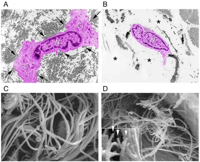
Fragmentation of collagen fibrils within dermis of aged/photoaged skin causes collapse of fibroblasts. A) Transmission electron micrograph of fibroblast (artificially colored pink for clarity) within dermis of young adult, sun-protected human skin. Note elongated, stretched appearance, and extended cytoplasm (C) away from nucleus (N) of fibroblast, which is in close proximity to abundant collagen fibrils (arrows, original magnification ×2,000). The top drawing of Figure 2 depicts a stretched fibroblast attached to intact collagen. B) Transmission electron micrograph of fibroblast (artificially colored for clarity) within dermis of photodamaged human skin. Note collapse of cytoplasm inward towards nucleus (N), and lack of adjacent collagen fibrils, compared to (A). Fibroblast is surrounded by amorphous material (*) The lower drawing of Figure 2 depicts a collapsed fibroblast surrounded by broken collgen. (C) Scanning electron micrograph of collagen fibrils in young adult human skin. Note that long fibrils are closely packed and fill space. Also note no apparent breaks in the fibrils (original magnification ×10,000). (D) Scanning electron micrograph (original magnification ×8,000) of collagen fibrils in photodamaged human skin. Note large gaps and numerous fragmented fibrils, compared to (C). Inset shows higher magnification of fragmented ends of fibrils (arrows, original magnification ×12,5000). Panels C and D are from Fligiel, S et al. Journal Investigative Dermatology, 120: 842-848, 2003.
