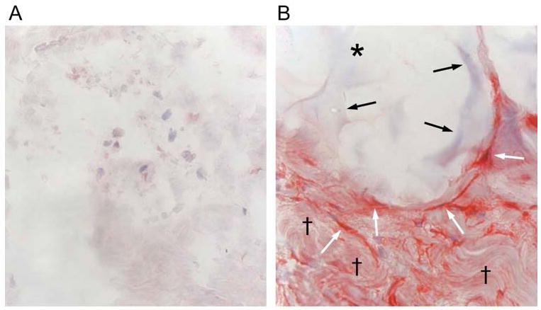
Figure 3.

Mechanical stretch induced by dermal injection of cross-linked hyaluronic acid filler stimulates collagen production in photodamaged human skin. (A) Saline vehicle (control) or (B) cross-linked hyaluronic acid filler (CLHA) was injected into photodamaged forearm skin. Skin biopsies were obtained four weeks post injection and analyzed for type I procollagen expression by immunohistochemistry. (A) Fibroblasts (nuclei stained blue) in saline-injected skin display barely-detectable procollagen expression (red stain). Also note amorphous space and fragmented thin appearance of collagen extracellular matrix (original magnification ×400). (B) In CLHA–injected skin, fibroblasts display intense type I procollagen immunostaining (red). CLHA appears as light blue material (black arrows) that occupies space adjacent to stretched fibroblasts (white arrows). Note also densely packed collagen fibrils (black daggers), not seen in saline-injected skin (A). These dense collagen fibrils are likely derived from CLHA-induced type I procollagen (i.e., conversion of type I procollagen into type I collagen) (original magnification ×400).
