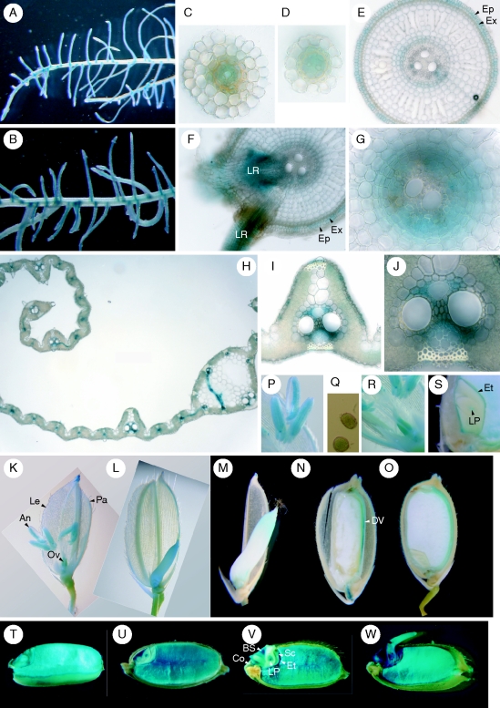
Fig. 2.
Cellular localization of IDEF2 expression as observed by histochemical staining of IDEF2 promoter-GUS expression in transgenic rice plants. (A) Fe-sufficient roots. (B) Fe-deficient roots. (C, D) Transverse sections of Fe-sufficient secondary roots. (E–G) Transverse sections of Fe-deficient main roots. (G) Enlarged parts of vascular tissues. (H–J) Transverse sections of Fe-deficient leaf blades. (J) An enlarged part of the phloem cells in (I). (K–S) Flowers and maturing seeds. (K, P–R) Before anthesis. (P) An enlarged part of the anther. (Q) Pollen. (R) Ovary. (L–O, S) After anthesis: 3 d (L), 5 d (M), 10 d (N) and 30 d (O) after fertilization. (S) An enlarged part of the embryo 30 d after fertilization. (T–W) Germinating seeds. Just before sowing (T), and 1 d (U), 2 d (V) and 3 d (W) after sowing. Abbreviations: An, anther; BS, bud scale; Co, coleorhiza; DV, dorsal vascular bundle; Ep, epidermis; Et, epithelium; Ex, exodermis; Le, lemma; LP, leaf primodium; LR, lateral root; Ov, ovary; Pa, palea; Sc, scutellum.

