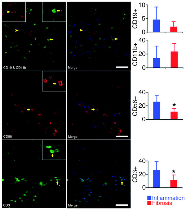
Figure 3.
Quantification of hepatic mononuclear cells in portal tracts. Immunofluorescence panels (left) identify the population of portal tracts by B lymphocytes (CD19), myeloid cells (neutrophils and macrophages: CD11b), NK cells (CD56), and T cells (CD3) in livers with a molecular signature of inflammation. Photos on the right depict the left photos after nuclear staining with DAPI (white bar = 50 μm). The graphs on the right show the average number (± standard deviation) of stained cells in portal tracts from six livers with the inflammation signature and from five with the fibrosis signature. *P < 0.05.

