Figure 5.
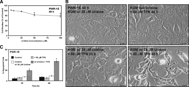
Uridine combined with TFN promotes cytotoxicity in PWR-1E cells. (A) The PWR-1E cells were cultured in KGM (control) or KGM with 5, 10, 25, 50, or 100 µM uridine for 2 days. The cells were harvested and counted as described in Figure 2A. The relative cell number in the uridine-treated cell populations is presented as a percentage of the untreated 48-hour control population. *P < .01 compared with the untreated control. (B) DIC micrographs showing PWR-1E cells cultured in KGMcontaining 25 µM uridine for 48 hours, the PWR-1E cells exposed to 50 µM TFN for 48 hours in KGM without uridine, and the PWR-1E cells exposed to 25 µM uridine and 50 µM TFN for 24 and 48 hours. Scale bars, 18 µm. (C) A summary of the hypoploid cells in the treatment populations described in panel (B). Me2SO was included in the control treatments without or with 25 µM uridine. *P < .001 compared with the TFN treatment alone.

