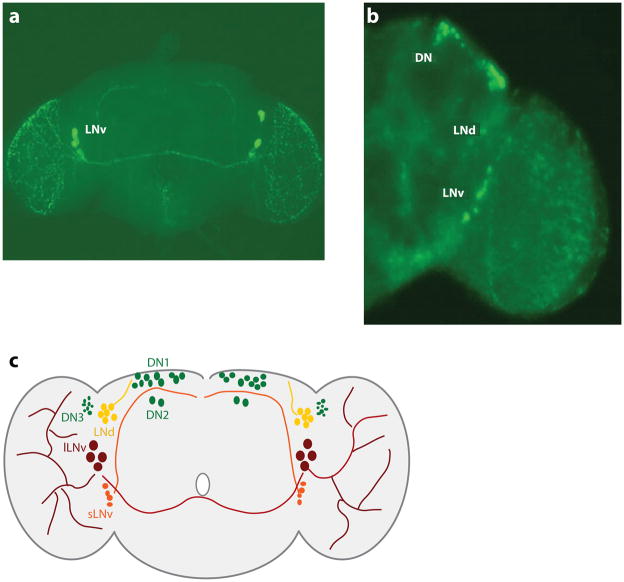
Figure 4.
The circadian pacemaker network in adult Drosophila brains. (a) Immunofluorescence of the circadian neuropeptide PIGMENT-DISPERSING FACTOR (PDF) illustrates the divergent projections stemming from ventral lateral neurons to other neural loci in the central nervous system. The small ventral lateral neurons (sLNv) send projections dorsally (toward the top of the image), whereas the large ventral lateral neurons (lLNv) send projections contralaterally and extensively into the optic lobes (on the right and left of the image). (b) tim in-situ hybridizations reveal the spatial orientation of pacemaker neurons in the central nervous system. Labels denote the location of the ventral lateral neuron (LNv), dorsal lateral neuron (LNd), and dorsal neuron (DN) clusters. Images in both panels a and b were provided by Ela Kula-Eversole. (c) Schematic of the Drosophila circadian pacemaker network. sLNv (orange) send projections dorsally. lLNv (maroon) send projections contralaterally and into the optic lobes (left and right sides of the schematic). LNd are indicated in yellow, and three groups of dorsal neurons are indicated in green.

