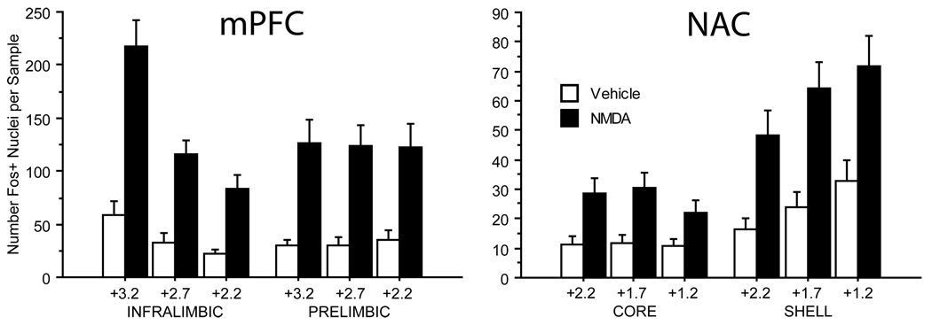
Figure 3.
Summary of c-Fos-protein expression for all unlesioned cases used in this study (n=30). In the mPFC (Left), bilateral VH MNDA infusion significantly elevated c-Fos expresssion in both the infralimbic and prelimbic cortices at all three coronal levels examined (see text for details). Whereas the amount of c-Fos expression did not differ between coronal levels in the prelimbic cortex, c-Fos expression in infralimbic cortex was significantly higher at the most rostral level (+3.2 from Bregma) compared to the other two levels (both p<0.0001). Similarly in the NAC (Right), bilateral VH NMDA infusion significantly elevated c-Fos-protein expression in both the core and shell of the NAC at all three coronal levels sampled in each (see text for details). Expression was found to be significantly more elevated in the shell compared to the core (p=0.0001), and in the shell there appeared to be a rostral to caudal gradient with c-Fos expression at the most caudal level (+1.2) significantly more elevated than at level +2.2 (p<0.01).

