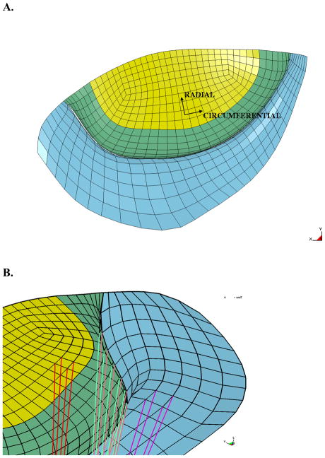
Figure 4.
Mitral valve in the undeformed configuration. A. Atrial view of the mitral valve with coordinate system defined. B. The underside of the mitral valve showing the chordal insertion points. Note the edge chordae attachments along the free-edge and the strut chordae attachments at the mid-section of the leaflets. The anterior leaflet is green and yellow, and the posterior leaflet is blue

