Figure 7.

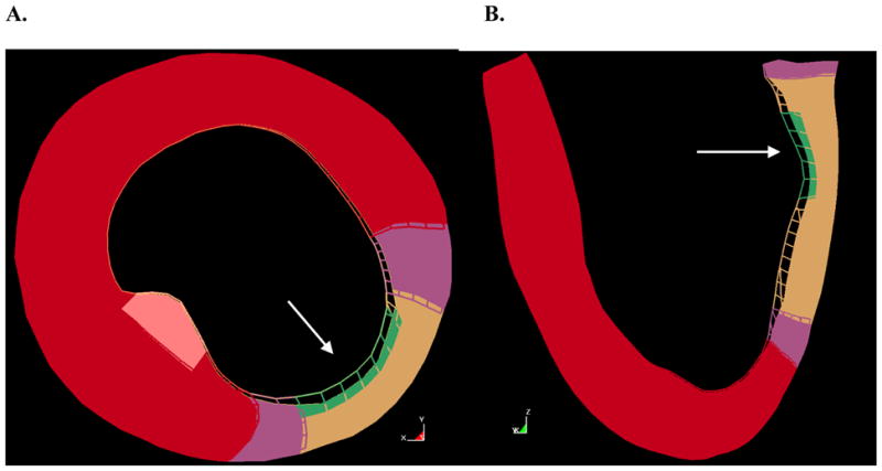
Overlay of stiff infarct model (wire mesh) and soft infarct model (solid) at end-systole. A. Short axis slice, B. long axis slice. The white arrow indicates the location of the posterior papillary muscle.

Overlay of stiff infarct model (wire mesh) and soft infarct model (solid) at end-systole. A. Short axis slice, B. long axis slice. The white arrow indicates the location of the posterior papillary muscle.