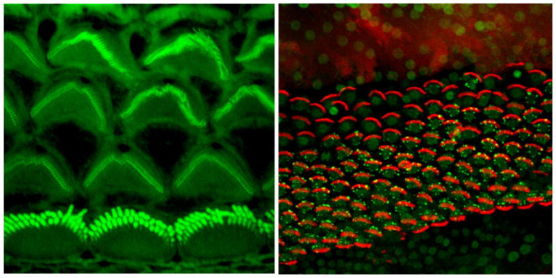
Figure 1.

Confocal micrographs of the luminal surface of the postnatal day 3 mouse organ of Cortiwhere actin is labeled with phalloidin (green) and of the regenerating mature chick basilar papilla where actin is labeled with phalloidin (red) and the early apoptosis marker TIAR is labeled with a specific antibody (green).
