
Figure 4. Injury and sirolimus alter p-S6RP expression in EC. The highest degree of S6RP phosphorylation in vivo occurs where EC are farthest from intact SMC.
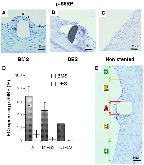
(A) p-S6RP expression was prominent in extracted porcine coronary arteries stented with BMS (arrows indicate positive staining cells in brown) and eliminated in sirolimus-eluting stents (B). Unstented control arteries (C) had an intact endothelium and media with no detectable p-S6RP staining. The incidence of p-S6RP positive EC in the luminal surface correlated with the nature of the underlying layer (D) and plotted by region relative to the stent strut (E). Region A is directly above the stent strut, B1 and B2 correspond to areas within the neointima adjacent to the stent strut, and C1 and C2 are regions where there is healthy endothelium, no detectable neointima and viable SMC within a normal medial layer (E). Only at a distance from the stent strut beyond the neointima/intact media boundary (zones C1 and C2) was the number of p-S6RP positive EC significantly reduced. n=12 (three experiments, four independent observations each).