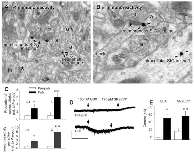
Fig. 1.
α4 and δ GABAA receptor subunit expression increases on dendritic spine membranes of CA1 hippocampal pyramidal cells at puberty. (A) α4 and (B) δ silver-intensified immunogold labeling (SIG) occurs along the plasma membrane of spines forming excitatory synapses. Shafts also exhibit immunoreactivity. Asterisks, postsynaptic density; t1 to t3, presynaptic axon terminals; a, nonsynaptic axons. In (B), the neck connects the labeled shaft with the spine. Scale bar, 500 nm. Arrowheads indicate SIG immunolabeling of the indicated GABAR subunits. (C) Proportion of labeled spines (upper panel: α4, *P < 0.018; δ, **P = 0.002) and immunoreactivity per spine (lower panel: α4, *P < 0.005, δ, **P = 0.00091) increase at puberty (Pub) relative to prepuberty (Pre-pub) (9). Error bars indicate SE of the mean. (D) Representative pyramidal-cell currents in response to the GABA agonist gaboxadol (applied continuously, arrows) and the GABAR antagonist SR95531 (applied continuously, arrows). Scale, 50 pA, 50 s. (E) Averaged data. *P = 0.0002, **P = 0.02, Pub versus Pre-pub.

