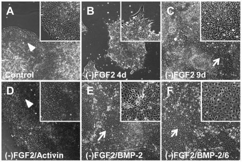
Figure 7. Morphological changes of hES cells after treatment with BMPs in FGF2-free mTeSR1.
H9 cells were split in regular mTeSR1 and changed to FGF2-free mTeSR1 24 h after splitting. After 4 days of growing in modified composition media, colonies were split in FGF2-free mTeSR1 and incubated for other 5 days (total of 9 days). Treatments with BMP-2 or BMP-2/6 at 100 ng/ml started 24 h after splitting and lasted for 4 days. Pictures were taken using an inverted microscope and 10X objective. A, control cells after 9 days of culture in mTeSR1. B, Incubation in FGF2-free medium ((-)FGF2) for 4 days. C, Incubation in (-)FGF2 for 9 days. D, Treatment with 100 ng/ml Activin A in (-)FGF2 for 9 days. E, Treatment with 100 ng/ml BMP-2 in (-)FGF2 for 4 days. F, Treatment with 100 ng/ml BMP-2/6 in (-)FGF2 for 4 days. Arrowheads indicate morphologically pluripotent cells; arrows point out morphologically differentiated areas. Insets belong to 3× magnification.

