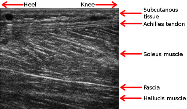
Figure 2.
An ultrasound image. The anatomic regions in the area around the Achilles tendon are explained by this example ultrasound image. All ultrasound images that occur in this article are captured at about the same anatomic region and are similar to this image. Case Example 1 and Case Example 2 are using a subset of such an image, covering only the Achilles tendon. Case Example 3 exploits the full image.

