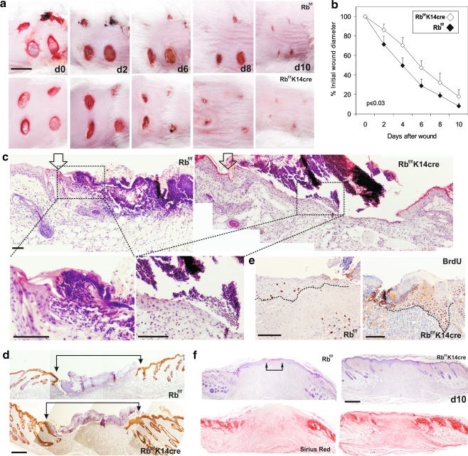
Fig. 7.
pRb ablation in epidermis accelerates skin wound healing. a Representative skin sections from control and Rb-deficient mice at different time points after wounding. Note the accelerated wound closure in mutant mice. Bar = 1 cm. b Plot summarizing the quantitative analysis of wound closure obtained from 4 different wounds induced in 4 different mice of each genotype. Data are shown as mean ± SE. c Haematoxylin-eosin stained sections of control and Rb-deficient mice showing the wound margins (denoted by arrows). Higher magnifications of the squared areas show the re-epithelialization front. d Keratin 5 immunostaining also demonstrates advanced re-epithelialization in pRb-deficient mice. Arrows denote initial wound margins. e BrdU-stained sections showing increased cell proliferation at the wound margins in Rb-deficient mice. Dashed lines denote the dermal-epidermal borders. F) Hematoxylin-eosin and sirius red staining of representative sections 10 days after wounding in control and Rb-deficient mice. The maturation of collagen fibers, denoted by intense red staining is slightly accelerated in Rb-deficient mice. Wound margins are still detected in control mice (arrows). Bars in c, d, e, f = 150 µm; Rbf/f, control mice; Rbf/f; K14Cre, Rb-deficient mice

