
Figure 5.
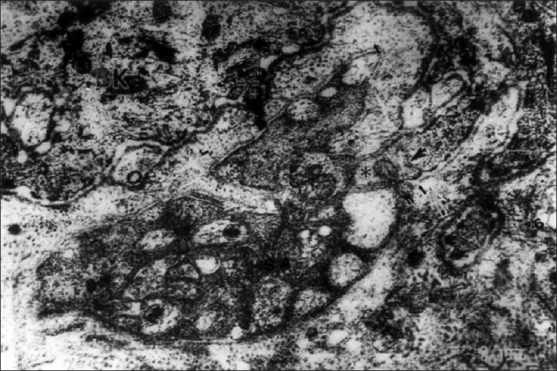
An intraepidermal SA has many axons, one which contact with the tip of microrete ridges (↑). Double arrows (↑↑) indicate the fusing portion between the basal lamina of SA and that of basal keratinocytes (EM ×20000)
Official websites use .gov
A
.gov website belongs to an official
government organization in the United States.
Secure .gov websites use HTTPS
A lock (
) or https:// means you've safely
connected to the .gov website. Share sensitive
information only on official, secure websites.
An intraepidermal SA has many axons, one which contact with the tip of microrete ridges (↑). Double arrows (↑↑) indicate the fusing portion between the basal lamina of SA and that of basal keratinocytes (EM ×20000)