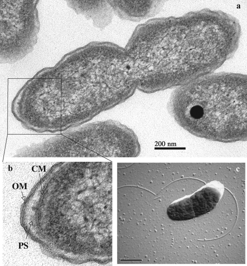
Fig. 1.
(a) Electron micrograph of a thin section of cells of strain EPR70T. (b) Details of the cell envelope of strain EPR70T. OM, Outer membrane; PS, periplasmic space; CM, cytoplasmic membrane. (c) Electron micrograph of a platinum-shadowed cell of strain EPR70T showing the presence of flagella. Bars, 200 nm (a) and 0.5 μm (c).

