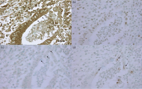
Fig. 6.
Representative photographs of immunohistochemical expression of TNF-α, IL-8, NF-κB p65 and phosphor-NF-κB p65 in semiserial sections of inflamed epithelium featuring cryptitis or crypt abscesses. (a) TNF-α expression in a crypt abscess. Note positive granules in the epithelial cytoplasm (arrows) (bar, 25 μm). (b) IL-8 expression in a crypt abscess. Note the positive reaction in the epithelial cytoplasm. (c) NF-κB p65 expression in epithelial nuclei in a crypt abscess (arrows). (d) Phosphor-NF-κB p65 expression in epithelial nuclei in a crypt abscess (arrows).

