Fig. 2.
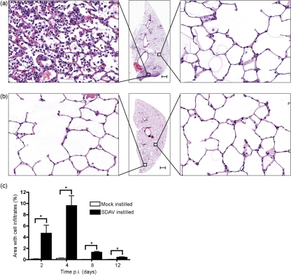
Lung pathology after SDAV infection. Sections of lung were stained with haematoxylin and eosin. (a) SDAV-instilled rats produced focal pulmonary lesions on day 4. (b) Mock-instilled rats did not have cellular infiltrates in alveoli. Bars in (a) and (b), 1 mm. (c) Whole slide images of haematoxylin and eosin slides were created using an automated scanning system (Aperio Technologies). Regions of the lung sections that had cellular infiltrates were scored by a pathologist. Asterisks indicate statistical significance (P<0.05) between SDAV and control samples. Error bars, sem.

