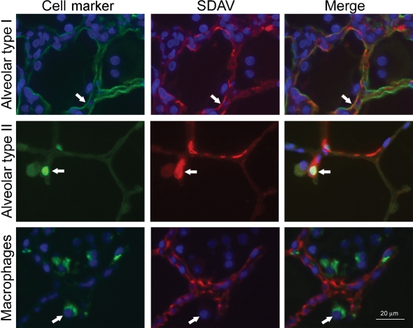
Fig. 4.
Identification of the cell types infected in the alveolar region. Rat lung paraffin sections were stained for cell markers (Alexa 488; green secondary antibody), SDAV (Alexa 594; red secondary antibody) and nuclei (DAPI; blue). Markers for alveolar epithelial type I cells (T1-α antibody), alveolar epithelial type II cells (TTF-1) and macrophages (rat CD68) were used to localize cells. SDAV was localized with an anti-MHV rabbit polyclonal antibody. Arrows indicate a cell identified by the marker antibody. Bar, 20 μm.

