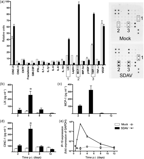
Fig. 5.
Cytokine expression by SDAV-infected rats. (a) BALF from SDAV- and mock-infected rats was analysed for cytokine and chemokine secretion using antibody arrays. Densitometry data were normalized to internal positive controls on each membrane (n=3) and graphed as relative units. Scans of autoradiographs for each membrane are shown to the right of the graph with relevant duplicate spots boxed with numbers corresponding to cytokines on the graph. (b–d) Cytokine levels for LIX, MCP-1 and CINC-1 were assayed in BALF using ELISA assays. All of the mock-instilled values for cytokine MCP-1 were below the level of detection. Asterisks indicate a statistically significant difference (P<0.05) compared with control samples. (e) Real-time quantitative PCR analysis of cytokine IP-10 expression. Error bars, sem.

