Fig. 5.
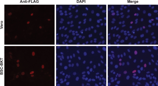
TruncTAg is localized to the nucleus. Vero (top panels) or BSC-BKT (bottom panels) cells were transfected with pcDNA3.1(+)-FLAGtruncTAg and analysed for localization of FLAG-tagged truncTAg by immunofluorescence, probing with an anti-FLAG epitope antibody as described in Methods. Representative fields of cells are shown at ×200 magnification. Anti-FLAG panels (left) are pseudocoloured in red; areas of colocalization with DAPI appear pink in the merged images (right).

