
Figure 23.
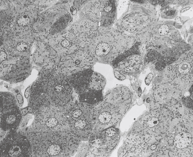
TEM micrograph of an injection-fixed human liver wedge biopsy at 700 × magnification, which shows the presence of dark and light cells next to each other. It is assumed that the differences in density reflect differences in fixation, which often occur in bad fixation.