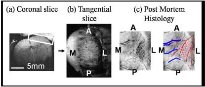
Fig. (1).
Experimental data orientations. Geometry is given in the anatomical reference frame. One representative animal shown. a) Coronal structural MR image (256*256) allows placement of b) a tangential slice covering the right hand cerebral hemisphere and hence surface of the somatosensory cortex (A-Anterior, P-Posterior, M-Medial and L-Lateral). BOLD and CBV activation maps superimposed onto such tangential sections were directly compared to post mortem histological sections (c). Photographic emulsion (previously injected into the vascular system) reveals the pattern of arteries (red) and veins (blue) in the somatosensory cortex. Individual barrels in the somatosensory cortex contain high levels of cytochrome oxidase. Staining for this enzyme allows direct visualisation of the whisker barrel field.

