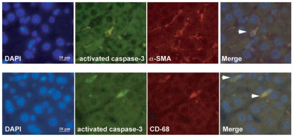
Fig. 6.

Immunofluorescent localization of activated caspase-3, α-SMA, and CD68 in livers of cirrhotic rats treated with JWH-133. Cell nuclei (blue), activated caspase-3 (green), α-SMA (red), and CD68 (red) fluorescent staining in cirrhotic livers treated with JWH-133 (1 mg/kg b.wt. for 9 days) was performed using 4,6-diamidino-2-phenylindole staining (DAPI) and specific antibodies. Colocalizations of activated caspase-3 with α-SMA (top) or CD68 (bottom) markers are shown in the merge panels (yellow; arrowheads) (n = 3). Original magnification, 400×.
