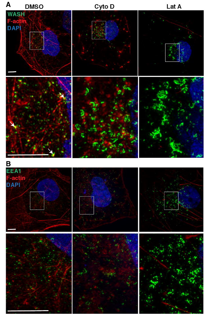
Figure 5.

Disruption of the actin cytoskeleton results in an enlarged and elongated WASH-containing and EEA1-positive endosomes. (A) WASH or (B) EEA1 visualized by immunofluorescence (green), F-actin stained with Alexa Fluor 568-phalloidin (red) and DNA stained with DAPI in COS7 cells treated with DMSO alone, or DMSO with 10 μM cytochalasin D (Cyto D), or 10 μM latrunculin A (Lat A). Imaging was by 3D structured illumination microscopy. Each panel is a representative maximum intensity projection of the 3D images. Actin colocalized with WASH structures (see arrows) in control (DMSO) treatment. Scale bars, 5 μm.
