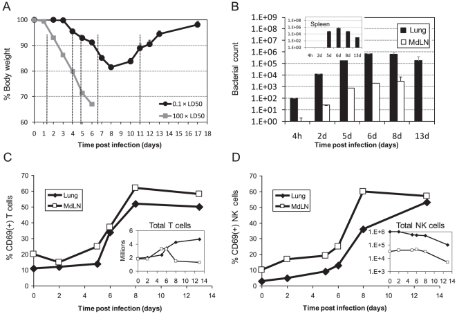
Figure 2. The effect of sub-lethal intranasal infection with 0.1×LD50 LVS on activation and recruitment kinetics of lymphocytes in the respiratory system.
(A) The mean weight of 60 animals in several independent experiments following intranasal infection with 0.1×LD50 LVS (black circles) or with 100×LD50 LVS (gray squares) is presented (Accumulation of six independent experiments with 10 mice in each experiment). Dashed vertical lines mark the time points selected for bacterial immunological analyses; (B) Live bacterial counts in the lungs (dark bars) and mediastinal lymph nodes (MdLN) (light bars) at the indicated time points after infection. Inset shows the concomitant live bacterial count in the spleen. The average counts out of three animals is shown in each time point; CD69 expression analysis on (C) gated T cells (NK1.1−CD3+ cells) and (D) gated NK cells (NK1.1+CD3− cells) in the lungs (closed diamonds) and MdLN (open squares) at the indicated time points after infection. The respective insets show the total numbers of NK or T cells. Cells were analyzed from three pooled animals at each time point. Panels B–D depicts a representative experiment out of three independent experiments performed. Each time point included three animals.

