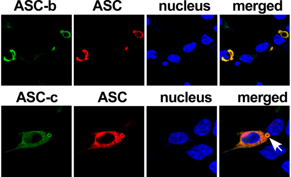
Figure 5.
Co-localization of ASC with ASC-b and ASC-c. HEK 293 cells were transiently co-transfected with HA-tagged ASC and myc-tagged ASC-b (1st panel) or ASC-c (2nd panel). Cells were fixed and immunostained with monoclonal anti-myc (Millipore) and polyclonal anti-HA (Abcam) antibodies, and Alexa Fluor-488 and -546 conjugated secondary antibodies (Invitrogen), respectively. Topro-3 was used to visualize the nucleus. All images were acquired using laser scanning confocal microscopy with a 100× oil-immersion objective. Panels from left to right show ASC-b/ASC-c (green), ASC (red), nucleus (blue) and a merged composite image. An arrow points to the aggregate.

