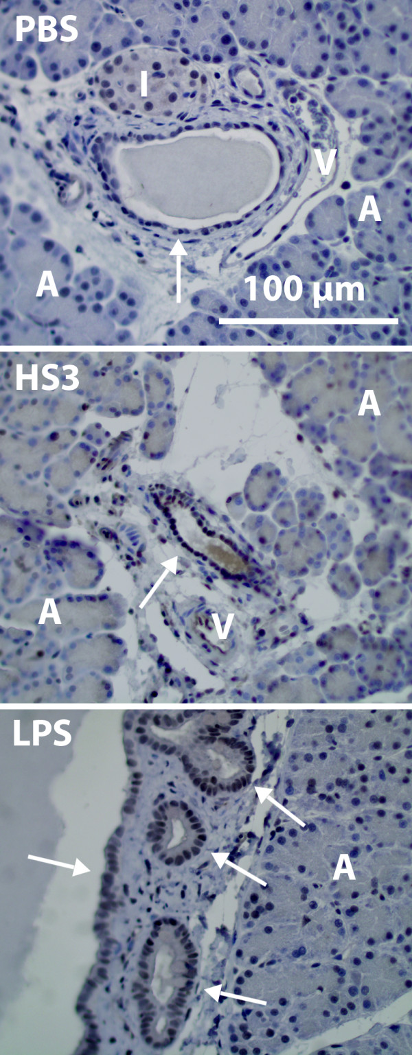
Figure 5.

MCP-1 expression 1 hour after infusion of PBS, HS and LPS. HS-infusion (middle) causes increased MCP-1 expression of the ductal epithelial cells (arrow) as compared to PBS control (top). Note that the different epithelial cells respond to different extent. LPS also induce a MCP-1 response of the epithelial cells (bottom). Other visible structures of the histological images include veins (V), islets of Langerhans (I) and acinar cells (A).
