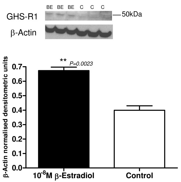
Figure 8.
Representative Western blot of GHS-R1 protein expression of β-Estradiol 10-8M (BE) treated hTERT-HM cells compared to control (C) untreated cells. The corresponding ACTB western blot is presented underneath the western blot. Molecular weights are indicated in kDa. Quantitative densitometric analysis of this western blot is presented, with β-Actin-normalised densitometric units for prepro-ghrelin plotted against treatment ± SEM (indicated with error bars). ** indicates a significance value of P < 0.01.

