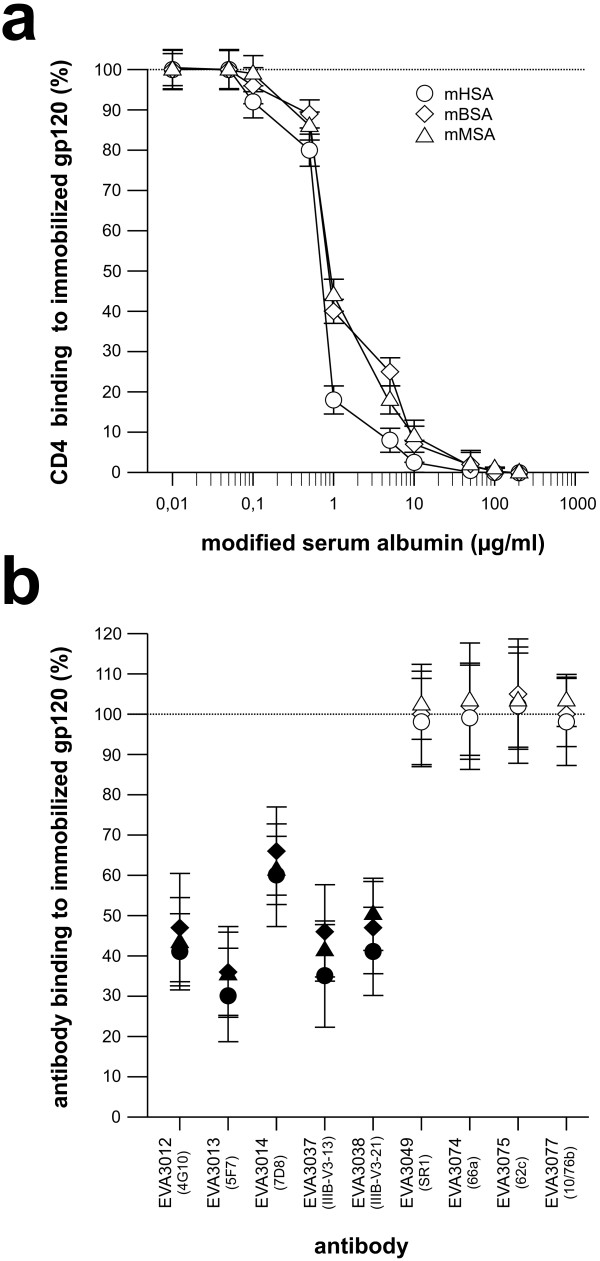
Figure 3.
Inhibition of CD4 and antibody binding to gp120. Binding of CD4 or antibody, in the presence of mHSA, mBSA and mMSA, to LAV gp120 was tested by ELISA. ELISA plates were coated with LAV gp120 (1 μg/ml, Protein Sciences Corporation) over night. After gp120 binding, the wells were coated using BSA (20 mg/ml). Modified serum albumin was added to the wells and (a) CD4 (1 μg/ml, Protein Sciences Corporation) or (b) antibody diluted in phosphate buffered saline (PBS) pH 7.5, was added 30 min later. After 1 hour, the plates were washed 5-times with PBS. Binding of CD4 to gp120 was carried out at various mSA concentrations. Gp120-bound CD4 was detected by a CD4 specific mouse monoclonal antibody (NCL-CD4-1F6, Novocastra) followed by a staining with an anti-mouse HRP-antibody conjugate (BioRad). Shown are the averages of triplicate wells. Binding of V2- and V3-loop-specific monoclonal antibodies (mab) was carried out in the presence of 10 μg/ml of mHSA. Gp120-bound antibody was detected using an anti-mouse-HRP-antibody conjugate (BioRad). Antibodies were designated according to the repository reference of the Centralized Facility for AIDS Reagents. In brackets: originators antibody designation. All measurements were carried out in triplicates. White symbols: V2 loop specific mab. Black symbols: V3 loop-specific mab.

