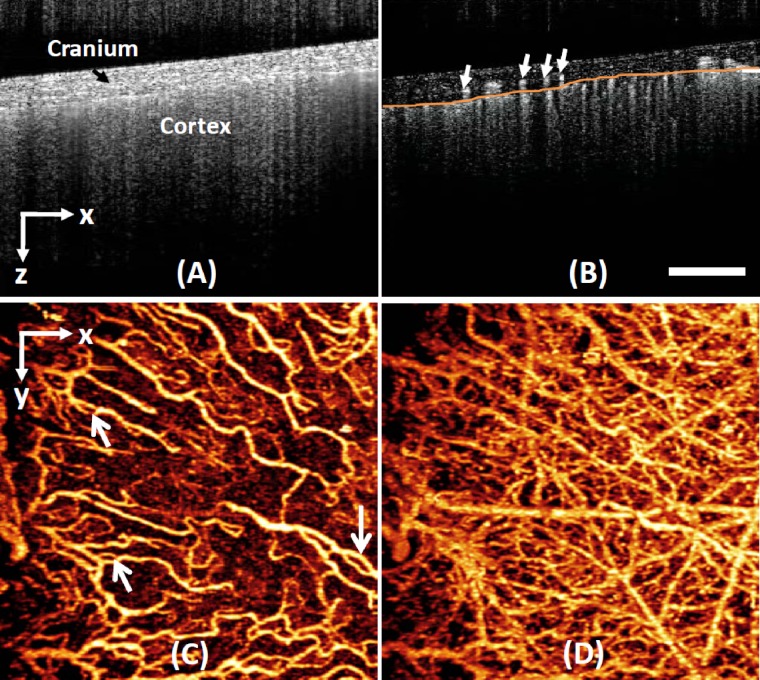
Figure 2.
Typical in vivo UHS-OMAG imaging of the microcirculation of the adult mouse brain with skull intact. (a) One typical UHS-OMAG cross sectional imaging (B scan) of microstructures showing morphological features, such as cranium and cortex, and (b) the corresponding depth-resolved blood flow image. The projection maps of blood flow network within (c) the meninges and (d) the cortex, respectively, are obtained from one 3-D scan. White bar=500 μm.

