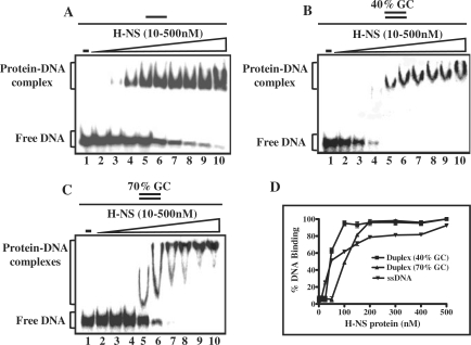
Figure 3.
Mycobaterium tuberculosis H-NS binds poorly to double-stranded DNA containing high GC base pairs. Reactions were performed with 5 nM of the indicated 32P-labeled DNA substrate in the absence (lane1) or presence of 10, 25, 50,100, 150, 200, 250, 300 or 500 nM H-NS (lanes 2–0), respectively. A single or two parallel lines on the top of each panel of the Figure denote single- or double-stranded DNA, respectively. The open triangle on the top of the gel image denotes increasing concentrations of H-NS. Reaction products were separated as described under ‘Experimental Procedures’ section. (A) ssDNA; (B) dsDNA (40% GC); (C) dsDNA (70% GC). The positions of free DNA and protein–DNA complexes are shown in the left-hand side of each panel. (D) Graphical representation of binding of H-NS to different DNA substrates. The extent of formation of H-NS–DNA complexes in (A–C) is plotted versus varying concentrations of H-NS. Error bars indicate SEM.

