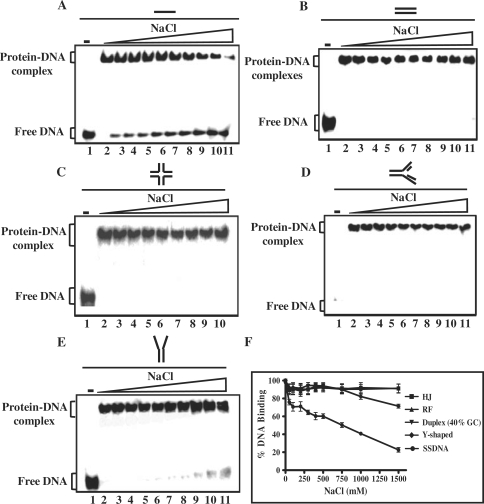
Figure 5.
Effect of NaCl on the stability of H-NS–DNA complexes. Reaction mixtures contained 5 nM of indicated 32P-labeled DNA substrate and 500 nM of M. tuberculosis H-NS. After incubation for 30 min, NaCl was added to the final concentration of 50 100, 200, 300, 400, 500, 750, 1000 or 1500 mM (lanes 3–11), respectively. After 10 min with NaCl, samples were electrophoresed on polyacrylamide gel, and this was followed by autoradiography as described under ‘Experimental Procedures’ section. (A) ssDNA; (B) dsDNA (40% GC); (C) HJ; (D) DNA replication fork; (E) Y-shaped junction; (F) the extent of dissociation of H-NS–DNA complex containing the indicated recombination intermediate is plotted versus varying concentrations of NaCl. Error bars indicate SEM.

