Figure 8.
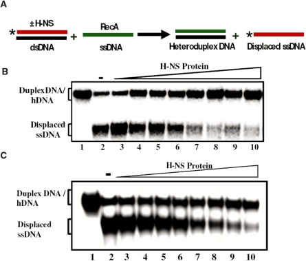
H-NS proteins suppress DNA strand exchange promoted by RecA protein. Assay was performed as described under ‘Experimental Procedures’ section. (A) Schematic depicting the experimental design. (B) Effect of M. tuberculosis H-NS on strand exchange by its cognate RecA. (C) Effect of E. coli H-NS on strand exchange by its cognate RecA. The positions of 32P-labeled displaced ssDNA, 32P-labeled duplex DNA or unlabeled heteroduplex DNA (hDNA) generated by RecA promoted strand transfer are shown in the left-hand side of the gel images. The open triangle on top of the gel images denotes increasing concentrations of H-NS. Lane1, control reactions lacking H-NS and RecA; lane 2, complete reaction in the absence of H-NS; lanes 3–10, complete reaction in the presence of 0.5, 1, 1.5, 2, 2.5, 3, 3.5 and 4 μM H-NS, respectively. An asterisk represents the labeled phosphate at the 5′ end.

