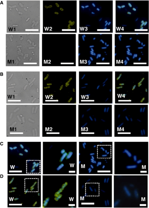
Figure 7.
Intracellular localization of the Dan protein. Wild-type E. coli KP7600 and its dan mutant JD24074 were grown in M9–0.4% glucose media for 24 h at 37°C under hypoxic conditions, and subjected to the indirect immuno-fluorescent microscopy according to the standard procedure described in ‘Materials and Methods’ section. (A) Wild-type strain KP7600 (W1–W4) and the dan mutant JD24074 (M1–M4) were grown under aerobic (A) or hypoxic (B) conditions. W1 and M1 represent; W2 and M2, Indirect immuno-blot against anti-Dan; W3 and M3, DAPI-staining; W4 and M4, merged images of DAPI and immuno-stained patterns. (C, D) The area shown by dotted square is expanded. Anti-Dan antibody was raised in rabbits using the purified Dan as an antigen. Cys3-labeled anti-rabbit IgG antibody was used as the secondary antibody. White bars indicate 5 μm.

