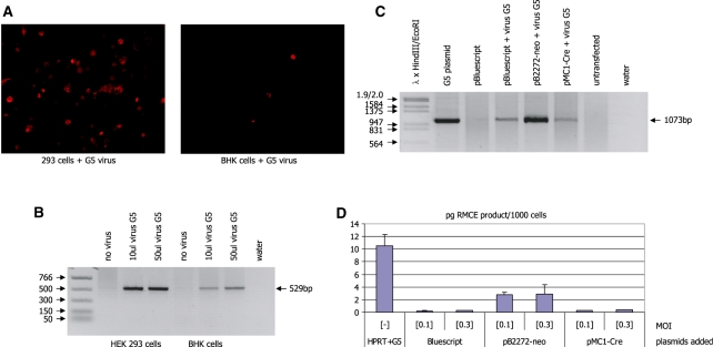
Figure 6.
(A) Red fluorescent staining in HEK 293 and BHK cells transfected with the plasmid pDSred-mito-2272-PGKneo and infected with the G5 adenovirus. Red staining of the mitochondria indicates the presence of the plasmid and the virus-derived Cre protein in the same cell. (B) PCR analysis of DNA derived from the cells in A using the primer combination DSred1R/CMVseq.1. Recombination between the two identical lox2272 sites in the plasmid, which is a prerequisite for the activation of the red fluorescent protein is indicated by the occurrence of a 529-bp band (marked by the arrow). (C) PCR analysis of DNA isolated from HEK293 cells transfected with the indicated plasmids and infected with adenovirus G5 at an MOI of 0.1. RMCE is detected using the primer pair pBKpA/neoint.4 which yields an indicative 1073 bp product (marked by the arrow). (D) Real-time PCR analysis of DNA isolated from HEK293 cells transfected with the indicated plasmids and infected with adenovirus G5 at an MOI of 0.1 or 0.3 (as indicated). RMCE is detected using the primer pair PGK5/CMVseq.1 (as in Figure 5) which yields an indicative 421-bp product. The amount of PCR product is shown as pg per 1000 cells.

