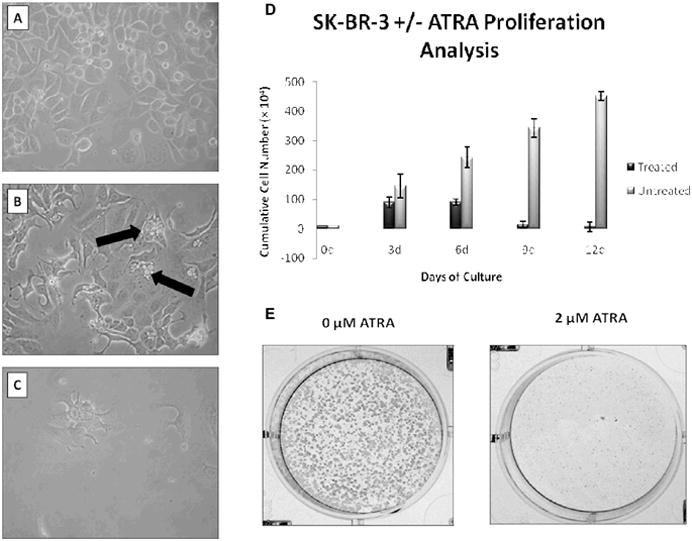
Figure 1.

Effect of ATRA exposure on morphology and proliferation in cultured breast cancer cells. Representative photographs, at 200X magnification of SK-BR-3 cells not exposed to ATRA (A) or exposed to ATRA for 6 (B) or 12 days (C). Black arrows in (B) indicate cells undergoing apoptosis. (D) Graphical representation of the means of cell counts taken during 12-day ATRA treatment. Means are +/− s.d. of triplicate experiments. (E) Representative SK-BR-3 cells colony formation.
