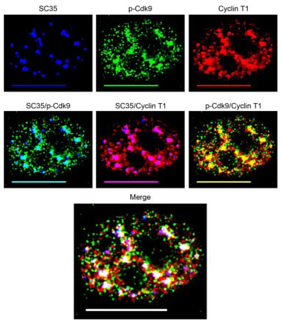
Fig. 2.
The active form of P-TEFb is located within nuclear speckles. HeLa cells were triple immunolabeled for SC35 (blue), p-Cdk9 (green), and Cyclin T1 (red) as shown in the upper parts. Co-localization of p-Cdk9 with SC35 (aqua), Cyclin T1 with SC35 (purple), and p-Cdk9 with Cyclin T1 (yellow) are shown in the middle parts. As can be seen, the large clusters of p-Cdk9 co-localize with nuclear speckles and to a high degree with Cyclin T1 (middle parts). The merge of all three channels shows that almost all of the active form of P-TEFb (p-Cdk9/Cyclin T1) is found within nuclear speckles. In the merge part areas of co-localization between p-Cdk9 and Cyclin T1 with nuclear speckles (SC35) appear as white, whereas those outside the speckle domains are indicated by yellow. Bar, 10 µm.

