Fig. 3.
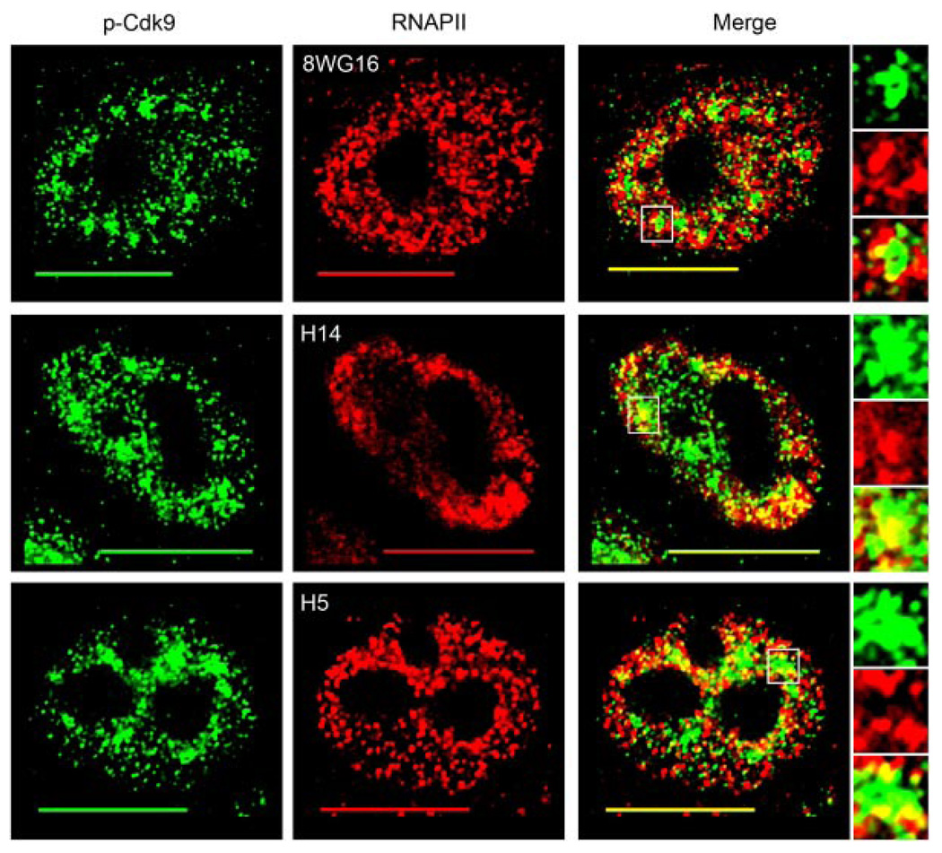
T-loop activated Cdk9 co-localizes mostly with the hyperphosphorylated forms of RNAPII. HeLa cells were dual immunolabeled for p-Cdk9 (green) and the various phosphoforms of RNAPII (red). Co-localization between p-Cdk9 and hypophosphorylated RNAPII (8WG16), Ser5-phosphorylated RNAPII (H14), and Ser2-phosphorylated RNAPII (H5) is shown in the upper, middle, and lower merge parts, respectively. Areas of co-localization are indicated by the yellow color in the merge parts and an enlargement of a speckle-like cluster of p-Cdk9 (boxed region in merge) from each cell is shown in the insets. Bar, 10 µm.

