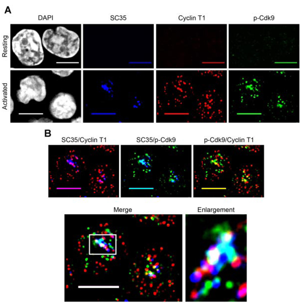
Fig. 7.
Active P-TEFb localizes to nuclear speckles in primary activated CD4+ T lymphocytes. A: Resting CD4+ T lymphocytes were cytospun onto glass coverslips and fixed immediately (upper parts) or activated (1 h) with PMA + ionomycin, cytospun onto glass coverslips and fixed (lower parts). The fixed cells were triple immunolabeled for SC35 (blue), Cyclin T1 (red), and p-Cdk9 (green), and the DNA was counterstained with DAPI to visualize nuclei. As can be seen, both the level of Cdk9 T-loop phosphorylation and the expression of Cyclin T1 and SC35 are highly up-regulated after 1 h of cell activation (compare upper to lower parts). B: Merge parts of the activated cells in A, showing the areas of co-localization between SC35 with Cyclin T1 (indicated by purple), SC35 with p-Cdk9 (indicated by aqua), Cyclin T1 with p-Cdk9 (indicated by yellow) and the co-localization of all three proteins (indicated by white). Ascan be seen, almost all of the active P-TEFb (CyclinT1/p-Cdk9) islocated either within or at the periphery of nuclear speckles in activated Tlymphocytes (merge part). This is particularly evident in the enlargement of a speckle region from one of these cells (boxed area on merge part) shown at the right of the merge part. The boxed region has been rotated 90° clockwise in the speckle enlargement for aesthetics. Bar, 5 µm.

