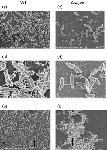
Fig. 2.
SEM images of biofilm formation by WT A. hydrophila SSU and its ΔahyRI mutant after 48 h cultivation at 37 °C on Thermanox coverslips stained with ruthenium red. (a, c) Compact aggregation; cells were well connected with filaments in biofilm formed by the WT strain. (b, d) Less aggregated cells in the ΔahyRI mutant biofilm, which were connected with fewer filaments compared with the biofilm formed by the WT bacteria. (e) A thick EPS was produced by the WT strain that was tightly bound to the surface of the cells (indicated by an arrow). (f) The ΔahyRI mutant produced a distinct type of EPS compared with that of the WT strain, and the EPS was loosely bound to the surface of the cells (indicated by an arrow).

