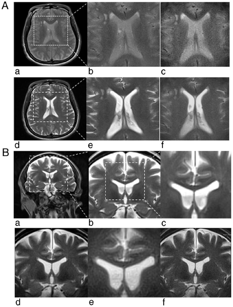
FIGURE 4.

A, Full FOV and 1/2 FOV PROPELLER brain images acquired without DEFT (a– c) and with DEFT (d–f). Full FOV (a) and inset from full FOV (×2) (b) PROPELLER images along with 1/2 FOV targeted-PROPELLER images (c) of the brain without DEFT showed reduced overall signal intensity and limited contrast between anatomic structures compared with those full FOV PROPELLER (d and e) and targeted-PROPELLER (f) images with DEFT. B, Full FOV (a) and insets from full FOV (×2 and ×4) (b and c) PROPELLER images of the brain compared with 1/2 FOV (d) and 1/4 FOV (e) targeted-PROPELLER images all with identical voxel sizes (0.8 × 0.8 mm2) but (d) and (e) requiring 1/2 and 1/4 the acquisition time of full FOV image (a). Half FOV targeted-PROPELLER image (f) with decreased voxel size (0.4 × 0.4 mm2) but same acquisition time as full FOV image (a).
