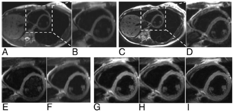
FIGURE 6.

Dark blood cardiac images acquired in short-axis orientation with HASTE (A with ×3 inset shown in B) and full FOV PROPELLER (C with ×3 inset shown in D) along with corresponding 1/3 FOV targeted-HASTE images (E and F with 6 averages) and targeted-PROPELLER (G, H with 60% k-space undersampling, and I with increased spatial resolution). Notice the blurring of HASTE images along the A-P PE direction, which is reduced with targeted-HASTE acquisition but inferior to isotropic resolution provided with PROPELLER approaches. Notice the superior depiction of fine anatomic structures within high resolution targeted-PROPELLER image (I) acquired within a single breath-hold.
