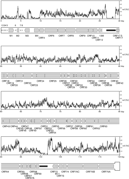
Fig. 2.
Variation between the genome sequences of WMHV and MuHV4. The lower part of the panels represents the genome, commencing in the first panel at the start of U and ending in the last panel with one copy of TR, which is shown in a thicker format. Protein-coding regions are depicted by shaded arrows, with connecting introns indicated by white horizontal bars and genes encoding the tRNA-like genes (1–8) shown as arrowheads. Internal tandem repeats are represented by black horizontal bars. The upper part of each panel shows the nucleotide divergence (nd) calculated for a 100 nt window, shifted by increments of 3 nt. A nucleotide position was counted as divergent if it differed between the two sequences; insertions and deletions were not scored.

