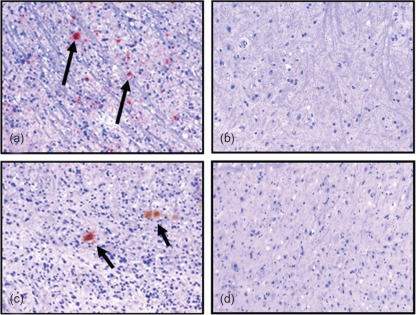
Fig. 3.
IHC from the obex region of the medulla from Tg(CerPrP) mice (×20 magnification). (a) PrPCWD (arrows) in a mouse exposed to CWD by aerosol versus (b) mouse exposed to sham inoculum. (c) Mouse exposed to CWD by IN route demonstrating PrPCWD aggregates (arrows) versus (d) mouse exposed intranasally to sham inoculum.

