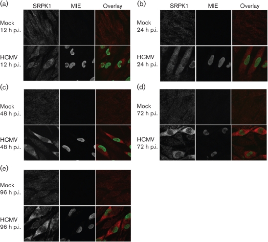
Fig. 1.
HCMV infection progressively increases the abundance of SRPK1 in the cytoplasm of infected G0-HFFs. G0-HFFs were HCMV infected (m.o.i.=3) or mock infected. At 12 (a), 24 (b), 48 (c), 72 (d) and 96 (e) h p.i., cells were harvested and sequentially incubated with anti-SRPK1 (red, 1 : 1000) and mAb 810 (specific for MIE, green, 1 : 500), followed by the corresponding secondary antibodies. Probed cells were imaged by confocal microscopy. Panels on the left and in the middle show greyscale imaging, whereas panels on the right show the merged colour images.

