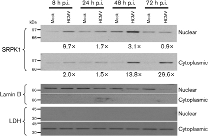
Fig. 2.
Subcellular distribution of SRPK1 in HCMV-infected G0-HFFs. HFFs (6.0×10⁷ cells) were growth arrested and mock infected or HCMV infected (m.o.i.=3), harvested at the indicated times, and fractionated into nuclear and cytoplasmic fractions. Fractionated proteins (20 μg) were resolved by electrophoresis, transferred to nitrocellulose and probed with anti-SRPK1 (1 : 1000), anti-lamin B (1 : 1000) and anti-LDH (1 : 1000). Probed blots were stripped of bound antibodies after exposure and reprobed with other antibodies. The fold inductions in SRPK1 abundance were determined by comparison of the densities of the bands from HCMV-infected cells divided by the densities of the corresponding mock-infected cells.

