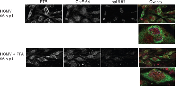
Fig. 5.
PFA treatment blocks HCMV-induced redistribution of PTB at late times of infection. G0-HFFs were HCMV infected (m.o.i.=10) and untreated or PFA treated as in Fig. 3. Cells were harvested at 96 h p.i. and stained for PTB (green), CstF-64 (red) and ppUL57 (blue) and imaged by confocal microscopy as in Fig. 4.

