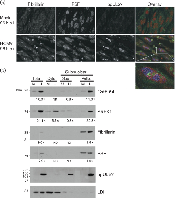
Fig. 7.
(a) HCMV infection induces the formation of fibrillarin-containing caps. G0-HFFs were mock infected or HCMV infected and harvested at 96 h p.i. as in Fig. 3. Cells were stained with anti-fibrillarin (green, 1 : 250), anti-PSF (red, 1 : 250) and anti-ppUL57 (blue, 1 : 250) antibodies and corresponding secondary antibodies. The left and middle panels are greyscale and the rightmost panels show all three channels merged. The bottom panel shows an enlarged HCMV-infected cell. (b) Co-sedimentation of cellular RNA-processing factors and nucleolar components with HCMV ppUL57, a VRC component. G0-HFFs were mock infected (M) or HCMV infected (H, m.o.i.=1) as in Fig. 2 and harvested at 96 h p.i. Cells were fractionated into cytoplasmic fraction, subnuclear pellet and supernatant (Sup) by modification of the nucleolar isolation procedure of Andersen et al. (2002). The cytoplasmic (Cyto) fraction was concentrated 5-fold and proteins (15 μg per well) were resolved by SDS-PAGE and blotted as described in Fig. 2. The blots were reacted with antibodies against CstF-64 (1 : 2000), SRPK1 (1 : 1000), fibrillarin (1 : 1000), PSF (1 : 2000), ppUL57 (1 : 500) or LDH (1 : 1000). Probed blots were stripped of bound primary and secondary antibodies after exposure and reprobed as required. The fold inductions in CstF-64, SRPK1, fibrillarin and PSF abundance were determined by comparison of the densities of the bands from HCMV-infected cells divided by the densities of the corresponding mock-infected cells as in Fig. 2 except where the mock- or HCMV-infected band was not detectable (nd).

